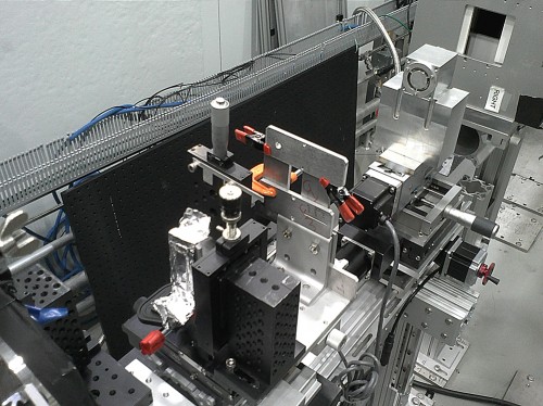
A team consisting of researchers from the Massachusetts Institute of Technology (MIT) and the National Aeronautics and Space Administration (NASA) is working to develop a microscope that uses beams of neutrons to produce high-resolution images. A series of papers published this year showed that the team’s proof-of-concept instrument is not only functional but also has significant potential to be improved [1].
Neutron-based microscopy provides a unique set of capabilities not offered in current types of microscopy, which utilize either light or electron beams. When neutrons pass through a material, strong interactions, which typically hold protons and neutrons together in the nucleus of an atom, can cause them to be scattered or absorbed into nuclei. This means that the image produced by passing neutron beams provides information regarding the neutron absorption of different sections of the specimen. Because neutron absorption is a property that varies across chemical elements, a neutron beam reveals the type, not density, of matter. This could prove immensely useful in studying soft matter, in which minute movements by hydrogen atoms can be detected as visible changes in neutron absorption [2].

Because neutrons do not have charge, they are not affected by electric fields. This makes them ideal for studying the interiors of electronic devices such as batteries and fuel cells, even while they are functioning [1]. However, although uncharged, neutrons do have spin and therefore a magnetic moment. Neutron microscopy is therefore also useful in determining the magnetic properties of materials. In particular, unaffected by electric fields, a neutron beam with a consistent direction of spin can allow magnetic properties to be isolated and studied, even within complex systems [2].
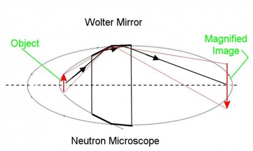
With all its interesting applications, neutron microscopes have been severely limited in the past by insufficient optics. Neutrons, unlike light beams, cannot be redirected using a simple mirror, as they would simply penetrate the reflective surface. The MIT/NASA team’s primary innovation involved utilizing a Wolter Mirror, which is typically used in X-ray microscopes, to focus neutron beams and magnify an image. The applied Wolter Mirror takes advantage of the fact that if a neutron hits a metal surface at a shallow enough angle, it will skid off. This allows a curved Wolter Mirror to redirect neutron beams [2].

To image the focused neutron image, the group used a film consisting of zinc sulfide overlaid with lithium. When the redirected neutrons contact the film, lithium atoms absorb the neutrons and undergo nuclear fission. This releases energy, which drives the zinc sulfide to glow. The light image can then be captured and analyzed [2].
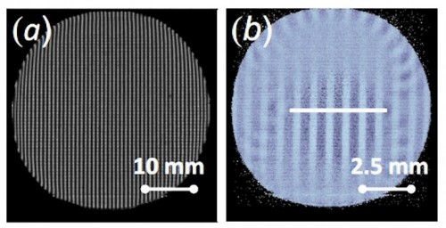
The team’s proof-of-concept instrument was able to achieve four-fold magnification, and they are currently working to optimize their design to achieve higher image resolution, magnification, and image brightness. It is estimated that the optimized prototype will cost several million dollars to produce [3]. However, this cost is minimal compared to the gains enabled by neutron microscopy.
With this research, a new type of instrument with immense potential is made possible. Because neutron microscopy will undoubtedly provide new perspectives in many fields of research, future work by this group is truly important.
Sources:
1. https://www.sciencedaily.com/releases/2013/10/131004125020.htm
2. https://www.gizmag.com/mit-neutron-microscope-reactor-prototype/29392/
3. https://web.mit.edu/newsoffice/2013/new-kind-of-microscope-uses-neutrons-1004.html
About the Author: Ben Gu is a sophomore in Berkeley College.
