Eyes on the prize, you jump into a river, furiously swimming for the shoreline. Yet, the second you reach for it, the current pulls you away. This is the challenge cancer drugs face in the human body: rapid clearance from the treatment site. The protection and safe delivery of these drugs as they travel to their target region are important factors in the drug’s success.
In order to combat this problem of rapid clearance, a group of Yale researchers has been studying therapeutic drug delivery through the use of sticky biodegradable nanoparticles. This technique targets tumors more efficiently by releasing drugs directly into the cancerous regions. The Yale study found that bioadhesive nanoparticles were capable of remaining in the tumor regions for long periods of time, demonstrating the potential for drug delivery through nanoparticles to fight cancer.
Promising potential
Surgery and chemotherapy are common treatments for aggressive tumors arising from the ovary and uterus. Unfortunately, many patients undergoing these therapies redevelop tumors or, in the case of chemotherapy, see their tumors become resistant to the treatment. For a little over the past decade, nanoparticles have emerged as a delivery system for agents such as drugs, offering targeting a larger portion of the drug to the tumor and causing less severe side effects. Nanoparticles can be engineered to enclose these drugs, protecting them on their way to the target site in the body.

Nanoparticle size plays an important role in the drug’s ability to stay in the region of the tumor. Too large a particle results in the drug’s accumulation in the lower abdomen, while too small of a particle results in a greater chance of abdominal fluid clearing the drug out of the system. With successful control of nanoparticle size, however, nanoparticles have the potential to allow for safer and more effective cancer treatment.
A sticky finding
Mark Saltzman, a professor at the Yale School of Engineering and Applied Science, is working to harness the potential of nanoparticles. His team developed nanoparticles with an outer coating of a polymer known as hyperbranched polyglycerol (HPG). HPG nanoparticles are like branched trees, where each branch is terminated in water-loving groups that make the particles water-soluble. This specific HPG outer coating has proven to be more effective than even the most highly regarded particle coating, possessing higher stability, lower risk of absorption in the body by proteins, and longer circulation in blood.
Experimenting with nanoparticles of different sizes, the team found nanoparticles measuring around 100 nanometers to be most effective in distributing throughout the body cavity and dispensing their encapsulated drug to the target site. The longer the time in the body, the longer the nanoparticles will release the therapeutic drugs into the tumors.
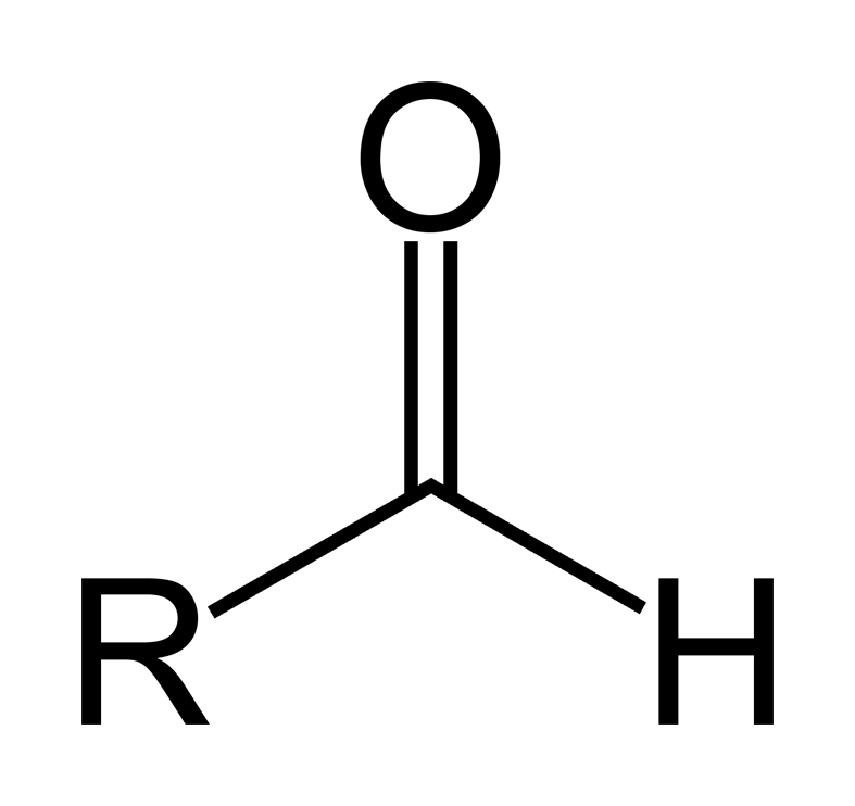
Initially, Saltzman and his laboratory team worked with non-sticky nanoparticles non-sticky that would circulate around the body for extended periods of time and eventually accumulate in the tumor. However, Yang Deng, a postdoctoral associate working in the laboratory, had an interesting finding. With organic chemistry techniques, he was able to make the nanoparticles that stick to protein-coated surfaces. This was done by using sodium periodate to transform the outer coating of the particles, generating aldehyde groups which are able to form bonds with other proteins.
Once the team discovered these sticky particles, the race was on to find suitable applications. The team was able to invent a new kind of sunblock that lasted longer on the skin, taking advantage of how the nanoparticles could stick to the skin’s top layer. However, the researchers also realized they could apply these sticky nanoparticles to areas such as cancer treatment. This is where Alessandro Santin came in.
Alessandro Santin, a professor at the Yale School of Medicine, treats patients with gynecological tumors with origins in the uterus and ovaries. In some cases, the tumor spreads out of the reproductive tract and into the abdomen, where it would grow in little clusters of cells that stick to the membrane surfaces of the abdomen.
Saltzman believed his sticky nanoparticles were relevant to Santin’s work for a variety of reasons. “If they’re sticky, we thought they would stick to the same membranes that the cancer cells stick to and they should get all over the abdomen, and eventually stick to the same surfaces tumor cells stick to,” Saltzman said. Saltzman and his team hypothesized that the sticky nanoparticles they had created could be applied to deliver drugs to tumors.
Targeting tumors with drugs

Saltzman, Santin, and their team sought to apply the their invention to cancer therapy. First, they tested how adhesive the sticky nanoparticles were compared to nonadhesive nanoparticles. To do this, they tested both particles in human umbilical cords, measuring how long they remained on the luminal surface. As a marker to quantify the level of adhesiveness, both particles were loaded with a dye, which emitted a fluorescent signal corresponding to the amount of particles in the region. The results showed that the bioadhesive nanoparticles had a longer signal time than that of their non-sticky counterparts. Now, the researchers could proceed with loading a drug into these nanoparticles.
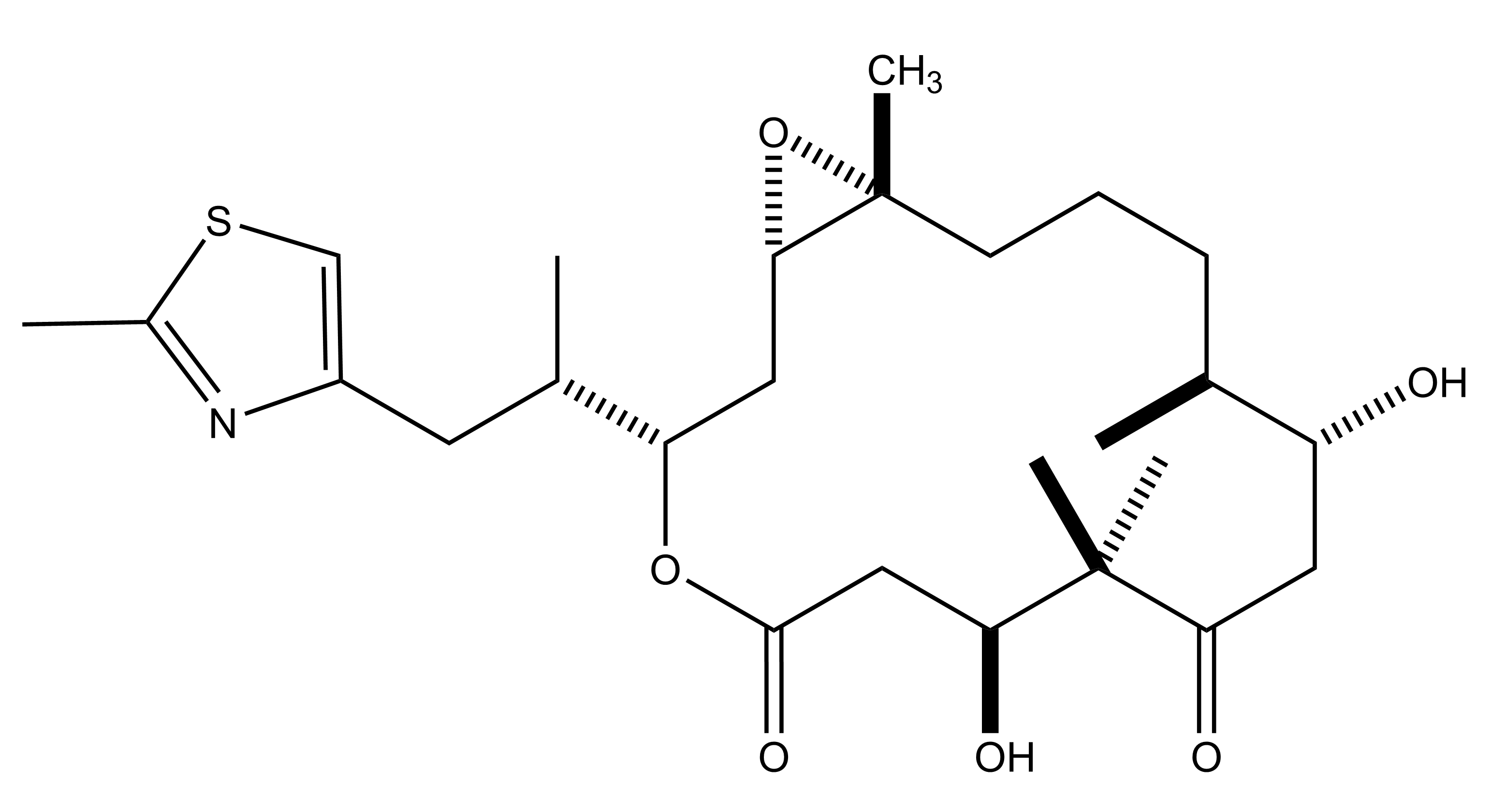
Epothilone B (EB), a drug that inhibits microtubule function, can prevent cancer cell division . The researchers chose to load EB into these sticky nanoparticles because of its efficacy against both ovarian and uterine cancers. However, the drug comes with its dangers. “The problem with this drug is that it’s so potent at killing cancer cells that it’s toxic,” Saltzman said. Therefore, a way to introduce the drug to the cancerous region gradually and more directly is crucial. Saltzman hypothesized that by injecting these EB-loaded bioadhesive nanoparticles into the space between the membranes that line the abdominal cavity, they could get the nanoparticles to form bonds with the proteins on the tissue surfaces. This would increase the length of time over which drug would be delivered to the tissue.
Traditionally, nanoparticles injected in this manner would be quickly cleared from the abdominal region. With the bioadhesive nanoparticles, however, the protein bonds formed with the cells layering this region held the nanoparticles in place. “We showed that if you take the particles and you expose them to a protein-coated surface, they stick very well,” Saltzman said. If successful, this would increase the bioavailabilty of the drug.
To test if this indeed happened, the researchers created a mouse model with human tumors in the abdominal region to simulate the environment in a human cancer patient. They loaded EB into the nanoparticle vectors and injected them into the mice. The results were promising — at the end of four months, the mice subjected to this treatment had a 60 percent survival rate, whereas only 10 percent of mice survived in the control groups. “We showed that by putting it in our bioadhesive nanoparticles, we can keep it in the abdomen for a long time with very little toxicity, particularly compared to the drug itself,” Saltzman said.

Sticking with it
The results of the Yale study offer the potential for more efficient methods for delivering drugs to target tumor cells. In the future, the researchers hope to increase the efficiency of drug delivery. For example, Santin is working on designing homing peptides that would target tumors specifically. “Doing so may increase the antitumor activity of the nanoparticle treatment,” Santin said.
Yet, more research needs to be done on the deliver system used in the study. Saltzman states that the researchers did not see a specific immune response when they observed general markers of immune activation, but that there are still more areas of concern to be addressed before the drug delivery system is ready to be used for cancer patient treatment. According to Santin, the researchers are working on an investigator-initiated trial using the drug. “This clinical trial will help to understand the potential of this novel agent in the treatment of chemotherapy-resistant ovarian cancer,” Santin said.
The results of the team’s work indicate the potential for bioadhesive nanoparticles as drug delivery agents to cancerous tumors. With more research on safer cancer treatment methods, nanoparticles may become a powerful weapon in our arsenal against devastating cancers.
Further Reading: Deng, Yang, Ediriwickrema, Asiri, Yang, Fan, Lewis, Julia, Girardi, Michael, Saltzman, W. Mark. 2015. “A sunblock based on bioadhesive nanoparticles.” Nature. 14:1278-1285. doi: 10.1038/NMAT4422.
About the Author: Jessica Trinh is a freshman and prospective biomedical engineering major in Branford. She enjoys volunteering for Synapse, Yale Scientific’s volunteer outreach program, being the Director of Operations for Resonance, Yale Scientific’s high school outreach program as well as the Communications Chair for Yale’s chapter of STEAM.
Acknowledgments: The author would like to thank Drs. Mark Saltzman and Alessandro Santin for their time and enthusiasm for sharing their research.
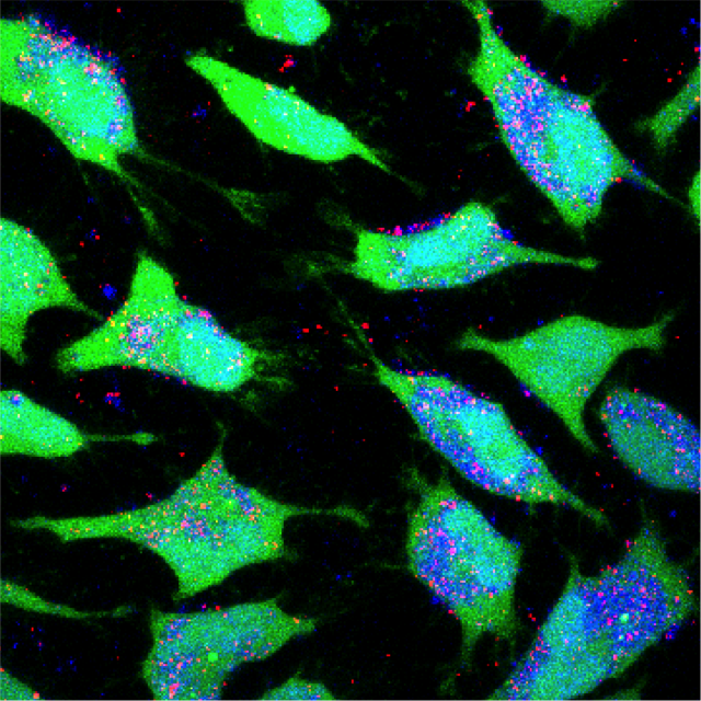
Cover image: Shown is an image of cancer cells (green) taking up fluorescently labeled nanoparticles (red), demonstrating the possibility of more efficient delivery of traditional cancer drugs. Image courtesy of Eric Song

