When pipelines rupture, water spews uncontrollably. Immediately, repairmen rush to the site, patching back up the damage. Similarly, such processes occur in the brain.
Intracerebral hemorrhage (ICH), a form of stroke, is characterized by rupturing blood vessels in the brain. Along with a mortality rate of 40-50%, there is no current treatment. Following the break in blood vessel, the battle is only half over. The immune system is activated, and macrophages such as microglia, a form of white blood cells, rush to the job gobbling up unwanted particles at the site of hemorrhage. However, once activated, the microglia respond in two possible ways. The classical response harms the brain by releasing pro-inflammatory molecules within the brain. The alternative response does just the opposite, releasing anti-inflammatory signaling factors aiding in brain tissue recovery. How such strikingly different responses could be activated in the human brain following ICH remains unknown.
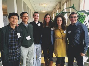
Lauren Sansing, associate professor in neurology at Yale University, along with Roslyn Taylor, lead author of a study and researcher in the Sansing lab, sought to examine the microglial response following ICH, and to determine the cause behind the activation of the alternative recovery responses. By studying changes in gene expression, Sansing and her team could pinpoint the mechanism behind these responses. To do this, the researchers analyzed the changes in the activation of 780 genes in microglia. They noticed that the most rapid changes in 78 of these genes occurred between days 3 and 7 following ICH. This time period, called the acute phase, is marked by edema—the swelling of the brain due to fluid. With hemorrhage causing damage to the brain, its microglia repair mechanisms are crucial. The researchers found that by day 7, microglia activate genes that aid in recovery, releasing anti-inflammatory molecules instead of pro-inflammatory ones. “It would make sense that microglia would turn off the inflammatory process pretty quickly,” Sansing said.

Once the researchers observed these changes in microglia phenotype, the question became determining what was mediating the alternative, reparative mechanism. Searching for possible pathways, researchers found a signaling protein most responsible: the TGF-β1 pathway. Earlier findings have shown the importance of this signaling pathway in the development of microglia. “It wasn’t surprising that TGF-β1 became important in this recovery phase, but it had never really been identified before,” Sansing said.
Now, they wanted to test whether introducing TGF-β1 protein would increase the repair functions of microglia. Taylor and her team studied the inflammatory responses in microglia in cell culture by treating them with thrombin, a protein-cutting enzyme, which stimulates microglia to react similarly to the response following ICH in the body. They then treated the cells with TGF-β1 protein and observed the response of microglia. Their results were promising—they found that inflammation was reduced after treatment.
Following their findings of the role of the TGF-β1 pathway in reducing inflammation in the classic microglial response, the researchers treated mice by injecting TGF-β1 protein directly into the brain and observed the microglial response. They found that TGF-β1-treatment given within 4 hours after the occurrence of ICH resulted in recovered motor function within a day. Researchers were then interested in how these results were relevant in human patients.
Applying these findings to human patients, the researchers found that the earlier the TGF-β1 pathway was induced, the higher the chance of recovery. Using the modified Rankin scale, which categorizes severity of stroke on a 7-point scale where 7 signifies death, the researchers measured the outcome of patients following ICH. Those with an earlier increased activation level of the TGF-β1 protein pathway 6 to 72 hours after ICH had less severe outcomes—and thereby a lower modified Ranking score—than those with later activation.
By studying the changes in response of microglia, Taylor and her team have provided promising outlook on the mechanism behind repair following brain hemorrhage. “I think that our findings will help provide further insights as to what TGF-B1 may be doing specifically in microglia in neuroinflammatory diseases,” Taylor said. While the researchers are still unsure what causes earlier activation of the TGF-β1 pathway in some patients, the results support the role of this pathway in repairing brain damage. Until then, Taylor and her team hopes other researchers will continue brainstorming ways in which the TGF-β1 pathway may be involved in therapeutic treatment for stroke victims. “The big question remains whether we can we actually intervene on this pathway by giving patients TGF-β1 protein,” Sansing said.
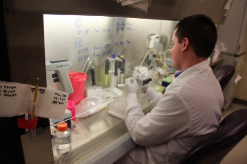
Featured Image: A member in the Sansing lab conducts experiments to study neurological diseases. Image courtesy of Jared Peralta.

