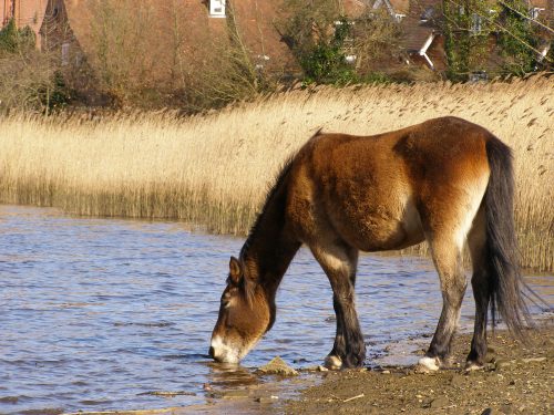
Water is essential for life. Its consumption is so intuitive, you probably don’t recall the last time you drank up. The body must work constantly to combat the water loss caused by urine production, sweating, and respiration, and it most often does this by inducing water intake. Though water regulation is an essential process, the brain mechanisms that govern thirst were unknown until recently. “Thirst is an ancient and extremely powerful motivational signal,” said Will Allen, a graduate student in the Department of Biology at Stanford, where he and his colleagues study the neural pathways that motivate thirst. Their results were reported in Science this September.
Animal behaviors, including thirst, are known to be motivated by two distinct mechanisms. The first is “drive reduction,” whereby thirsty animals learn to drink to avoid the “aversive state” of thirst. The second mechanism works the opposite way: thirsty animals have greater incentive than their satiated counterparts to drink for the “reward” of satiety. Previous studies suggested that the latter reward mechanism is more likely in the case of thirst, but could not provide neural mechanisms to support this hypothesis.
The study at Stanford provided evidence that thirst is an “aversive internal state” and is actually driven by the first mechanism. “This study provides a neural implementation of a long-discarded theory from psychology about how motivational drives produce behavior,” Allen said. The conclusion was enabled by recently-developed techniques that allowed researchers to selectively access, activate and manipulate neurons in the brain.
To identify the specific group of neurons that encode thirst, the researchers altered the genetic sequences of mice so that during thirst, cells would fluoresce in red, via a fluorescence gene aptly named tdTomato. By comparing tdTomato fluorescence between water-deprived and water-satiated mice, they were able to pick out the cells that had increased activity due to thirst.
The researchers then isolated the tdTomato-positive cells and used RNA sequencing to determine individual genetic expression profiles. Based on these profiles, the researchers identified two important cell types. One of these cell types was concentrated in the median preoptic nucleus (MnPO)—a brain region in the hypothalamus involved in osmoregulation, thermoregulation, and sleep homeostasis. Since the hypothalamus is known to regulate many metabolic processes, and the MnPO is involved in electrolyte balance, these neurons were expected to be the primary motivators of thirst.
The researchers hypothesized that if MnPO neurons indeed motivate thirst, then artificially activating them should induce water consumption. They inserted genetic “switches” into mice MnPO neurons to turn thirst on and off with a laser beam and a fiber optic implant. Photoactivation of the neurons in water-satiated animals induced water consumption, and the rate of water consumption scaled with frequency of stimulation. Water-deprived mice were trained to press a lever to obtain water, such that photoactivation of the neurons induced lever-pressing.
To determine if the activity of these neurons causes an aversive state, two experiments were performed. First, mice were put into a chamber, one half of which photoactivated the neurons. Mice invariably learned to avoid that half. In the second experiment, water-deprived mice were provided a lever that turned off photoactivation. They learned to vigorously lever-press, even though no water was provided upon pressing. Moreover, the higher the frequency of photoactivation, the higher rate of lever-pressing. Their response indicated that higher levels of MnPO neuron activity are more aversive and result in stronger thirst.
Finally, the researchers investigated how MnPO neuron activity changes upon water intake. They proposed three mechanisms: first, that neuron activity decreases due to the anticipation of water; second, that neuron activity decreases as water is consumed; and third, that neuron activity continues until a certain satiety threshold is reached, after which it abruptly turns off. Once again, they inserted the tdTomato fluorescence gene into MnPO neurons, allowing the neurons to fluoresce with an intensity proportional to their activity. When mice were allowed to press a lever to obtain water, their MnPO activity decreased gradually over several minutes, much more slowly than during free drinking. Once neuron activity reached a minimum, the mice stopped lever-pressing. This indicated that MnPO neurons receive quantitative real-time feedback from the body, and adjust their activity accordingly.
The study shows how MnPO neurons regulate animals’ water-seeking behavior, improving our understanding of how thirst is motivated. It remains to be seen, however, whether the alternative reward mechanism has a role in thirst, how MnPO neurons know when satiety is achieved, and what makes up the secondary pathways that enable a coordinated response, supporting the survival of animals for hundreds of thousands of years.

