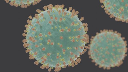
While Moderna and Pfizer’s mRNA vaccines prevent sickness with COVID-19, they do not help people who have already become sick with the novel coronavirus. These prophylactic measures target the most famous protein encoded by the SARS-CoV-2 genome: the spear-shaped spike protein that transverses the viral protein coat and punctures the host cell, allowing the virus to inject its genetic information. The spike protein enables the infection to occur, but what is it about the novel coronavirus that poisons our cells and makes us sick?
SARS-CoV-2—like many other viruses such as Influenza A and SARS-CoV, the agent responsible for the SARS outbreak in 2003—contains an mRNA transcript that encodes its viral proteins. Among them is nonstructural protein 1, or Nsp1. As its name would suggest, nonstructural protein 1 is not a structural building block of the viral particle. The main function of the viral protein Nsp1 is to stop the host cell from expressing its own genes. Produced early after viral infection, this protein starts reshaping the cellular environment to accommodate viral proliferation. A previous study found that, in SARS-CoV, Nsp1 is also necessary for viral replication, making it a vital component of sickness progression and a strong candidate for target therapeutics against coronaviruses.
With the COVID-19 outbreak, coronavirus research became critical, and scientists applied what was already known about SARS-CoV to make hypotheses about the novel coronavirus. Yale Molecular Biophysics and Biochemistry professor Yong Xiong, whose research group studies how viruses suppress and escape a host’s immune system, hypothesized that SARS-CoV-2 Nsp1 is likely critical for disease progression and poisoning host cells.
To test this hypothesis, his collaborator, associate professor Sidi Chen, investigated twenty-seven of the twenty-nine proteins encoded by the SARS-CoV-2 genome. Chen transfected each protein individually into human lung epithelial cells and found that out of the twenty-seven SARS-CoV-2 proteins tested, Nsp1 caused the most severe decrease in cell viability. To confirm that Nsp1 is the linchpin of this phenotype, a new population of cells was transfected with a mutated, defunct copy of Nsp1. This group of cells remained healthy, leading Xiong and Chen to conclude in a recent paper published in Molecular Cell that SARS-CoV-2 Nsp1 is “one of the most potent pathogenicity protein factors of SARS-CoV-2 in human cells of lung origin.”
Shifting gears
After Xiong and his collaborators knew with greater certainty what leads to pathogenicity, they began to investigate how Nsp1 led to this cell sickness. Nsp1 infection causes a large-scale shift in the host cell’s transcriptome, with the expression of 9,262 genes being altered as a result of this protein’s presence in the cell. By sequencing cellular mRNAs and quantifying the amount of each mRNA transcript present using mRNA-seq, the research team was able to determine which host genes were affected by Nsp1 expression. Nsp1 expression led to the decreased expression of 5,394 genes, the majority of which are related to protein synthesis, cellular metabolism, and the immune system. To express the proteins encoded in their own genome, cells need the protein-production machinery, the ribosome, and energy to translate their mRNA transcripts into proteins. By suppressing genes involved in these processes, Nsp1 shuts down cellular protein synthesis—hijacking the host cell, re-routing resources to build viral machinery, and dampening the cell’s immune response to allow the infection to occur.
The connection between Nsp1 expression and the genes it upregulates is less clear than those it downregulates. Nsp1 upregulates the expression of 3,868 genes that encode transcription factors that regulate higher-order chromatin structure, homeobox genes that are most known for driving body patterning, DEAD-box genes that regulate RNA metabolism, and regulators that drive cell fate determination. How upregulation of these genes might affect the pathogenicity of SARS-CoV-2 is not yet understood. “Logically, Nsp1 programs the cellular transcriptome in order to redirect cellular resources to the virus, but there is nothing specific that jumps out to us,” Xiong said. How Nsp1 alters gene expression on a molecular level is still unclear as Nsp1 has no nuclear activity, meaning that it never enters the host cell nucleus where all the cell’s genetic information is stored.
The two-pronged approach
In the case of SARS-CoV, Nsp1 has been shown to bind to 40S, the small ribosomal subunit, to block translation of mRNA into protein and promote cleavage and degradation of cellular mRNA. However, the molecular mechanisms of these activities remained unexplained. Recent advancements in cryogenic electron microscopy (cryo-EM) and Xiong’s role in bringing this technology to Yale has made it possible to use these clues from SARS-CoV to look at SARS-CoV-2 activities at the atomic scale.
Xiong used cryo-EM to investigate how Nsp1 inhibits protein synthesis. By freezing proteins down to cryogenic temperatures (approximately below negative 150 degrees Celsius), Xiong was able to capture proteins in their native form and image these native structures at the resolution of 2.7 angstroms, about the width of a water molecule. His lab found that the C-terminus, or back end, of the Nsp1 protein tightly binds to the mRNA entry channel on the 40S subunit, while the N-terminus interacts more loosely with subunit’s head domain. “Think of a body with a neck and head. Around the neck is the mRNA path, where it is loaded and translated,” Ivan Lomakin, an associate research scientist in the Bunick lab and expert in human protein synthesis, explained. “Part of Nsp1 binds to this path. The other portion binds to the head, which is a moving part that would otherwise enable mRNA to slide along the channel.” While the C-terminus of Nsp1 physically sits in the entry channel at the neck and binds to the ribosomal RNA and ribosomal proteins uS3 and uS5, the rest of the Nsp1 molecule interacts with the head domain of the ribosome.
The exact effect of this is unknown since the N-terminus does not bind tightly to the ribosome, so the cryo-EM image could not precisely determine how the N-terminus makes contact with the 40S subunit. Nsp1 also competes with some initiation factors critical for eukaryotic translation for binding to the 40S subunit and locks the 40S subunit in a “closed” conformation, which is the state where the ribosome is unable to load mRNA.
In addition to preventing mRNA from loading onto the ribosome, previous studies focusing on SARS-CoV have shown that Nsp1 prompts the cutting of host cell mRNA. mRNA stability is determined by many structural features within the mRNA transcript, which contains caps, tails, and sequences that can loop back on themselves and provide stability. By prompting cutting of the mRNA transcript, Nsp1 targets the host cell transcript for rapid degradation. How Nsp1 does this remains a completely open question.
How does SARS-CoV-2 mRNA escape?
“The two-pronged approach on inhibiting cellular protein production is just half the story. The other half is how viral mRNA escapes,” said Xiong. With such a well-defined notion of Nsp1 blocking and cutting host cell mRNA, an important question remained: how does the viral mRNA escape this mechanism and translate its own genome?
Xiong explains that the answer likely lies in the 5’ untranslated region (UTR) of the SARS-CoV-2 genome, a portion of the mRNA strand upstream of the protein-encoding segments. “We have a clue from the literature already. Some viruses harbor a mutation that prevents mRNA cutting,” he said. These genes harbor an internal ribosome entry site (IRES) that directly binds to the 40S subunit and enables protein translation initiation without the normally required 5’ mRNA cap and cellular initiation factors.
Previous studies found that SARS-CoV relies on its 5′-UTR for evading the Nsp-1-mediated translation block. SARS-CoV-2 may use an “IRES-like” mechanism where the 5′-UTR enables translation without the initiation factors blocked by Nsp1 binding to the ribosome. In addition, viral 5′-UTR could cause the Nsp1 C-terminus to dissociate from the mRNA entry channel of the 40S subunit, effectively unplugging the protein from the channel and allowing the ribosome complex to form and to load mRNA. However, the exact mechanism by which SARS-CoV-2 evades the translation shutdown still remains to be demonstrated.
Nsp1 as a therapeutic target
The new COVID-19 vaccines are designed to prevent us from getting sick with COVID-19, but they do not cure the many who already are sick. Moreover, there is insufficient information as to whether these vaccines guard against other related coronaviruses.
Scientists already speculate that, like the flu vaccine, we may need booster shots regularly to protect against evolving SARS-CoV-2 strains. Although coronaviruses do not mutate as rapidly as the flu, there are already multiple new and more infectious forms of the novel coronavirus, and COVID-19 is the third coronavirus outbreak to occur in the last two decades.
Unfortunately, it seems that coronaviruses will not be leaving the human population soon, and therefore it is critical that, in addition to vaccination prevention, there are also effective treatment options. Nsp1 is a particularly attractive target due to its largest role, among all viral proteins, in affecting cell viability.
Xiong’s research on how Nsp1 leads to pathogenicity through host cell translation suppression suggests it may be an effective therapeutic target. “Our hope is that what we learn from these interactions will give us something on the treatment front,” Xiong said. While more research needs to be done on the molecular interactions between Nsp1, the ribosome, and the viral mRNA transcript before therapies can begin to be developed, Nsp1 seems to be a promising future drug target.
Acknowledgements
Thank you to professor Yong Xiong and Ivan Lomakin for their time and commitment to their research.
About the Author
Britt Bistis is a senior majoring in Molecular Biophysics and Biochemistry. She works in the Noonan lab investigating how mutations in high-confidence autism risk gene CHD8 alter corticogenesis and the regulatory mechanisms through which this may occur. Outside of the lab she can be found doing volunteer work in programs for special needs students and science outreach programs or horseback riding.
References
Yuan, S., Peng, L., Park, J. J., Hu, Y., Devarkar, S. C., Dong, M. B., Shen, Q., Wu, S., Chen, S., Lomakin, I. B., & Xiong, Y. (2020). Nonstructural Protein 1 of SARS-CoV-2 Is a Potent Pathogenicity Factor Redirecting Host Protein Synthesis Machinery toward Viral RNA. Molecular cell, 80(6), 1055–1066.e6. https://doi.org/10.1016/j.molcel.2020.10.034
Wathelet, M. G., Orr, M., Frieman, M. B., & Baric, R. S. (2007). Severe acute respiratory syndrome coronavirus evades antiviral signaling: role of nsp1 and rational design of an attenuated strain. Journal of virology, 81(21), 11620–11633. https://doi.org/10.1128/JVI.00702-07.
Kamitani, W., Huang, C., Narayanan, K., Lokugamage, K.G., Makino, S. (2009). A two-pronged strategy to suppress host protein synthesis by SARS coronavirus Nsp1 protein. Nat Struct Mol Biol, 16(11), 1134-1140. DOI: 10.1038/nsmb.1680

