
Art by William Archacki
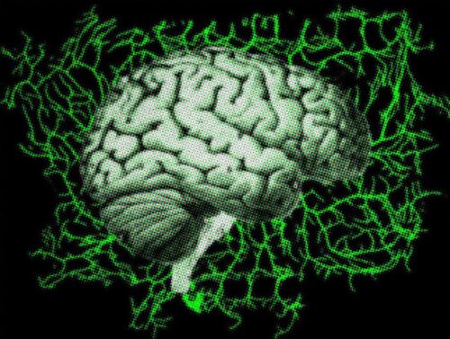
Nature loves branches. Look closely, and you’ll see branching structures everywhere—not just in trees and shrubs, but also in the tentacles of coral, in the blood vessels of mammals’ circulatory systems, and even within the human brain. Branches known as dendrites enable signaling within the brain by stretching outward from every neuron, forming intricate message-moving structures that look something like roots on a forest floor.
Dendrites are difficult to observe. For a scientist hoping to understand the structure of dendrites, the first problem is the complex three-dimensional geometry which translates poorly to a two-dimensional image from a microscope. The second problem is the simple fact that dendrite growth occurs inside sophisticated organisms which are difficult to observe under a microscope alive. New research out of Yale’s Quantitative Biology Institute (QBio) sought to overcome these obstacles and accurately track the growth patterns of dendrites, producing models that could explain the mechanisms behind dendrite growth.
The team at QBio led by Professor Joe Howard focused on certain sensory neurons that grow just under the skin of fruit fly larvae. The fruit fly neurons they studied are relatively flat and remain visible through the skin when tagged with a fluorescent pigment. Sonal Shree, an associate research scientist at the Howard lab who worked on the project, noted that fruit flies also have the advantage of quick maturation. “They have a very complex morphology, and they attain this morphology in days”, Shree said. In comparison, the development of the human nervous system takes place over months and years.
Once the researchers were able to image living dendrites under a microscope, they needed to determine the rules that guide dendrites through development. From a single image, it’s difficult to tell how dendrites form their intricate structures. “One of our main concerns was whether these neurons are growing by dynamics, like branching, or just inflating like a balloon with the growing larva”, Shree said.
Through time-lapse photography of the same neurons over a period of 120 hours, the researchers identified patterns in how new branches form and the directions they travel. They confirmed that dendrites truly do grow by branching rather than inflation, and they found that development occurs in cyclical periods of growth, pause, and shrinking.
“To explore the space, the best strategy is to grow and see the neighborhood, to check it out, and then grow again,” said Sabyasachi Sutradhar, a physicist at QBio who was responsible for the computational modeling. “There are other cells which are chemically attracted through directed growth or guided growth, but if you don’t include that, this is a very general framework by which neuron cells can grow.”
The team at the Howard lab hopes that the quantitative models they created to analyze dendrite data can be used to analyze other branching structures in organic systems. They hope to study the dynamics behind the growth of other thin, complex structures like the microtubules that scaffold and fortify cells’ structures.