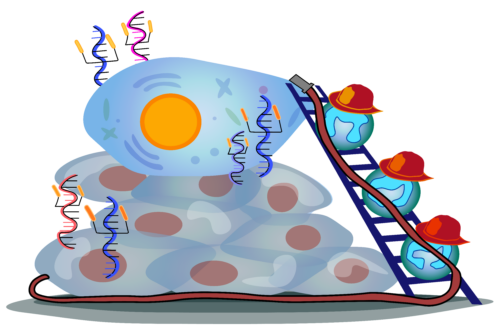
Art Courtesy of Luna Aguilar.
Ribonucleic acids (RNA) are among the most important molecules in the human body. Early in the evolution of life, RNA controlled all molecular and cellular functions, including the catalysis of biochemical reactions. Today, RNA carries out myriad functions within cells, such as communicating genetic information from DNA to functional protein products, regulating gene expression, and defending cells against viral infections. Recently, however, it has been discovered that RNA is not only confined within the cell but also present on the cell’s surface.
RNA…Outside of Cells?
In 2011, RNA was first discovered binding to the cell membrane—which separates a cell’s interior from the outside—in bacteria in the lab of Ronald Breaker, a Sterling Professor of Molecular, Cellular, and Developmental Biology at Yale. It was not until 2020 that Liangfang Zhang and Sheng Zhong, professors of bioengineering at the University of California, San Diego, discovered what they coined “membrane-associated extracellular RNAs.” These “maxRNAs” were RNAs stably associated with the outer membrane surface of human cells, presenting a mysterious new role of RNAs. This discovery was quickly compounded upon by Nobel Laureate Carolyn Bertozzi in a 2021 paper announcing the discovery of “glycoRNAs,” small extracellular noncoding RNAs with special sugar molecules attached to them. These recent discoveries opened the door for further exploration into the functions of extracellular RNAs.
When Jun Lu, an associate professor of genetics at the Yale School of Medicine (YSM), learned of these findings, he was surprised by the reported stability of the extracellular RNAs. “Outside of cells, we know that there are lots of RNases that act as Pac-Mans to chew up extracellular RNAs very efficiently,” Lu said. Indeed, the idea that RNAs could exist outside of cells for prolonged periods posed a confounding mystery. Interested in uncovering the functional roles of glycoRNAs, Lu approached his upstairs lab neighbor, frequent collaborator, and expert on neutrophil biology, Dianqing (Dan) Wu, the Gladys Phillips Crofoot Professor of Pharmacology at YSM. The result was a study affirming the presence of extracellular RNAs on neutrophils and their function in facilitating neutrophil recruitment to infection sites.
The Firefighters of the Blood Stream
Neutrophils are the most common white blood cell, or infection-fighting cell, in the human bloodstream. While extensive research has been done on their immune function at infection sites, less is known about how they navigate to these locations. Traditionally, neutrophil biologists have focused on the process of rolling adhesion as an important step for this movement. Neutrophils typically float in the bloodstream untethered to any scaffolding. But when injury or inflammation exposes the cell adhesion molecules on endothelial tissue lining blood vessels, neutrophils adhere to the walls of blood vessels and roll toward the site of injury to fight accumulating pathogens.
Before this collaboration, Wu didn’t have much experience in RNA research. Being a neutrophil biologist, however, he did have the intuition that something in the neutrophil-endothelial interaction included sugar-binding. Neutrophil biologists had previously identified a class of proteins found on endothelial cells called selectins that appeared to bind proteins with attached sugars on the surface of neutrophils. These interactions were reversible, allowing neutrophils to swiftly engage with endothelial tissue repeatedly, which underlies the ability of these cells to roll along blood vessel walls.
To investigate the presence of cell surface RNAs in this process, the authors utilized a sophisticated technique called click chemistry, which won Bertozzi her Nobel Prize in Chemistry in 2022. Click chemistry describes reactions that are simple, highly efficient, and selective, just like clicking a button. Ideally, these reactions should also avoid side reactions and provide high yields of the desired product at mild conditions. Click chemistry is often used to join molecular building blocks together with applications in bioconjugation, materials science, and drug discovery. In this study specifically, the authors employed a bioorthogonal reaction, which is a click reaction in a biochemical setting that selectively targets biological molecules without disturbing other cellular processes.
First, the authors labeled the RNA of cells with a sialic acid sugar mimetic called Ac4ManNAz, which acts as a molecular tag, forming glycoRNAs. Then, using click chemistry, they attached a biotin molecule to the labeled sugars in the glycoRNAs. The attached biotin subsequently served as a beacon, allowing glycoRNAs to be easily detectable. The researchers found a strong biotin signal on the surface of neutrophils, suggesting the startling presence of glycoRNA. They then confirmed that glycoRNAs bind to a specific selectin called P-selectin on endothelial cells. By binding P-selectin, glycoRNAs help facilitate the neutrophil-endothelial interaction necessary for neutrophils to roll along blood vessels.
The combination of discovering the glycosylated ligands on the neutrophil cell surface and elucidating at least one functional role of extracellular RNA involved a fair bit of serendipity. “I think both of us were a little skeptical initially [at the magnitude of the effects of removal of extracellular RNAs]. We wanted to make sure this wasn’t just a single-time interaction,” Wu said. Together, Wu and Lu’s labs were able to demonstrate the functional importance of neutrophil cell surface RNAs dozens of times, giving them the green light in terms of reproducibility.
How Firefighters Fight the Fire
While much remains unknown about how neutrophils produce extracellular glycoRNA, biologists know that neutrophils aren’t recruited until they are needed. When tissue is injured or inflamed, it must somehow signal for neutrophils to come and bind to the tissue. Lu likens this process of neutrophil recruitment to firefighters rushing to a fire. Upon injury, damaged cells release proinflammatory proteins called cytokines that signal nearby endothelial cells to express selectins, like P-selectin, which are normally tightly controlled. This “fire” prompts the neutrophils, acting as firefighters, to attend to the site of injury. “Selectins are like glue that try to capture circulating white blood cells—mostly neutrophils because of their abundance—which starts the neutrophil infiltration process,” Wu said. P-selectin–neutrophil–glycoRNA binding is central to this glue-like interaction, but this is likely just part of the picture.
Wu and Lu performed an experiment called RNA sequencing on glycoRNA isolated using click chemistry from three kinds of neutrophils in the mouse bloodstream and several human cell lines. Analyzing the mapping of these RNAs to the mouse or human genomes indicated specific rules that could govern RNA glycosylation. Lu describes these rules as a “licensing step.” Although the existence of these rules is not currently known, Lu speculates that they may involve special RNA sequences, structures, or modifications. Wu expressed that it may not be sufficient for an RNA to be glycosylated for recognition by the P-selectin; P-selectin’s specificity may include the RNA as well. These discoveries are only the first steps in characterizing the specificity of the P-selectin-neutrophil-glycoRNA interaction, but the methodology used in this study will likely inform future explorations of extracellular protein-glycoRNA interactions across the body.
How did glycoRNAs get outside of the cell?
The authors proposed two potential models to explain the mechanism by which glycoRNAs moved from the cytoplasm inside the cell to the outer surface of neutrophils. In the cell-to-cell model, cellular RNAs are released from one cell and are captured on an adjacent cell’s surface. On the other hand, in the cell-autonomous model, the production and transport of cellular RNAs to the cell surface occur in the same cell. To differentiate between these models, a co-culture experiment was conducted. One group of neutrophils was labeled with the Ac4ManNAz sugar and a fluorescent green dye, while another group was only labeled with a fluorescent red dye. These cells were then mixed and incubated together. Afterward, the researchers found only strong fluorescent signals in the green cells but not the red cells, confirming the second cell-autonomous model of the production and transportation of glycoRNAs across the cell membrane.
Once they had confirmed the cellular origin of glycoRNAs, Lu and Wu began to consider the pathways by which glycoRNAs could leave the cells. One such pathway was inspired by the C. elegans worm. In this model organism, the Sidt1 gene was found to encode RNA transporters that facilitated the uptake of digested RNA in the gut into the cells of the worm across cellular membranes. Therefore, the Yale scientists reasoned that the Sidt genes expressed in neutrophils could be facilitating the transport of RNAs across the cell membrane. To test this hypothesis, they disrupted the expression of both Sidt1 and Sidt2 in cells in a knockdown experiment. As a result, the presence of Ac4ManNAz-labeled glycoRNAs was abolished, highlighting the crucial role of Sidt RNA transporters in the presence of glycoRNAs in cells. Importantly, the Sidt-knockdown cells also exhibited a significant reduction in in vivo recruitment to inflammatory sites, underscoring the essential role of Sidt genes in the functionality of neutrophils.
The Future of the RNA World
This novel collaboration between a neutrophil biologist and an RNA biologist is only the beginning of a growing field focused on glycoRNA interactions outside the cell. Lu and Wu are both eager to continue their collaboration and begin answering the many questions opened by this paper. As this field is still in its infancy, Lu suggests that it will take some work to even begin elucidating the initial mechanistic questions, such as exploring the molecular pathways involved in making extracellular glycosylated RNAs and figuring out the environments in which they are selectively produced or glycosylated. “We can’t work on all of these questions, so we have to be careful about picking the lower-hanging fruits first to work on. Then, I expect many people will start to work on it,” Lu said. After that, Wu and Lu hope other labs explore more disease-specific questions, like studying the disease conditions in which these RNAs are dysregulated and whether there are any therapeutic or diagnostic roles for extracellular RNAs.

