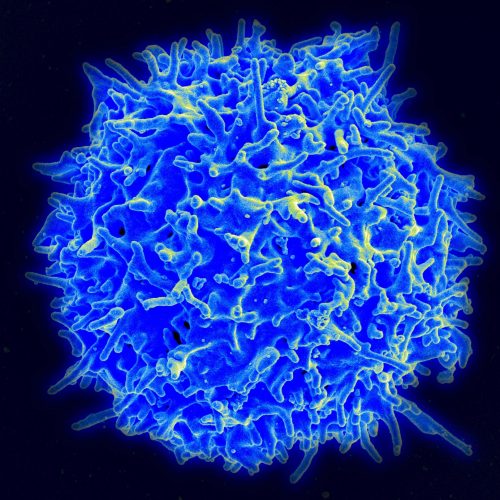
The human immune system is widely studied due to its integral role in health, from preventing infection to the development of autoimmune diseases. At Yale University, Professor Anna Pyle’s lab is specifically interested in the function of innate immune receptor retinoic acid-inducible gene I, or RIG-I, which serves as a first line of defense against infection. RIG-I, among other similar receptors, is responsible for detecting viral RNA or damage and alerting the rest of the immune system. However, mutations in RIG-I can lead to over-activation and excessive signaling, causing individuals to be more susceptible to autoimmune disorders. Because of this, researchers in the Pyle lab are interested in studying the activation of the domain in RIG-I responsible for signaling.
When designing an experiment to study the conformational changes of the protein in the signaling pathway, the Pyle team ran into several issues with methods currently used to study similar pathways. According to Thayne Dickey, a former postdoc in the Pyle lab, a big problem was that “cells are messy.” In vivo studies in molecular biology face a myriad of challenges due to the dynamic nature of cells and interdependence of many molecules and their functions. Because of the complications of in vivo studies, researchers in Pyle’s lab spearheaded the effort to create an in vitro method to study RIG-I in order to streamline analysis of its activation.
The team of researchers was particularly interested in how RIG-I becomes activated to eject its signaling domain and how incorrect activation leads to autoimmune disorders. The researchers turned to a technique called Florescence Resonance Energy Transfer, or FRET. This assay involves labeling a protein in two places with different fluorophores, in which the florescence of one fluorophore excites the other fluorophore. “FRET tells us the distance between two positions in the protein, so if it changes structure we can see that by a change in fluorescence,” Dickey explained. FRET has previously been used in the field of drug discovery because it enables scientists to test millions of potential drugs, offering a rapid fluorescent output that shows which compounds have the desired effect. Despite the prevalence of FRET in drug discovery, it is rarely used in vitro when studying purified proteins and conformational changes. “The possibility of extending fluorescent analysis…to RIG-I analysis was attractive,” Dickey recalled. When comparing this assay to other similar options, Dickey said that FRET stands out because of its high throughput and reliability.
One of the largest challenges the researchers encountered next was getting an efficient label for the protein of interest. Dickey revealed that trial and error was necessary to figure out how to stick the fluorophores onto the protein. “The conjugation site had a dozen constructs,” Dickey said. In some constructs, the protein would not be labeled correctly, so the whole assay would fail. It was critical for protein labeling to accurately measure the ejection of the RIG-I signaling domain because it is the first step in receptor activation.
Finally, with a working assay that revealed conformational changes of RIG-I before and after activation, the team analyzed the role of ATP and RNA binding in the signaling pathway. Previously, ATP was assumed to play an integral role in the ejection of the signaling domain of RIG-I and was required for full activation of RIG-I. However, after running the FRET assay, the researchers found that ATP only has a small effect on the conformation and ejection of the signaling domain. It changed the way Dickey thought about RIG-I since “[this experiment] shows that RNA is sufficient to [activate RIG-I] on its own and ATP is not actually required for the activation of RIG-I.” While there is still some uncertainty around the role of ATP, the lab found that RNA binding alone caused a reduction in FRET consistent with the signaling domain being ejected from the protein. Taking it a step further, this result implies that RNA binding is the key biophysical step in the activation of RIG-I that could be targeted by drugs.
Another key result of this study was the reversible property of RIG-I signaling domain ejection. When the binding RNA was digested with nuclease, the FRET measurement increased back to the levels of the initial inactive state of the protein. This finding demonstrated that RIG-I can be deactivated and returned to its inactive state after the signaling domain has been ejected. The use of the FRET assay was essential to discovering these conformational changes in the protein. The discoveries of the Pyle lab present the opportunity for RIG-I to be a potential therapeutic target for blocking autoimmune disorders. Already, the lab is diving into applications of RIG-I activation and all it can offer the field of immunotherapy.

